Human Heart
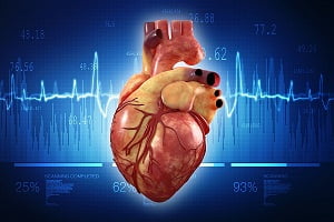
The heart is a muscular organ about the size of a closed fist that functions as our body’s circulatory pump. It takes in deoxygenated blood through the veins and delivers it to the lungs for oxygenation and then pumps this oxygenated blood into the arteries. The arteries provide oxygen and nutrients to bodily tissues by transporting the blood throughout the body.
Location of heart, Size of heart
Human heart is located in the thoracic cavity medial to the lungs and just behind and slightly left of the breastbone.
The human heart is precisely situated at the left side of your chest. Its size is approximately the size of your fist when closed. The exact position of your heart is between the left and right lungs. It is behind the breastbone and slightly inclined towards the left.
If you keep your right hand in the central part of your chest, and move it slightly to the left, you can reach the exact location where your heart resides. The spine is behind the heart and lungs are located on either sides of your heart.
More: Heart Rate By Age: What Is A Good Heart Rate As You Age?
More: What Are Abnormal Heart Sounds (Heart Murmurs)?
Anatomy of the Heart (Heart Structure)
Our heart consists of several layers of a tough muscular wall called the myocardium. A thin layer of tissue (called pericardium) covers the outside. Another layer called endocardium covers the inside. The heart cavity is divided into four chambers. The upper chamber is called an atrium (or auricle) and the lower chamber is called a ventricle. There are two atria that act as receiving chambers for blood entering the heart, and two ventricles that pump the blood out of the heart.
Chambers of the heart
The heart has four chambers, as discussed herein.
- The right atrium: It receives blood from your veins and pumps it to the right ventricle
- The right ventricle: It receives blood from the right atrium and pumps it to your lungs.
- The left atrium: It receives oxygenated blood from the lungs and pumps it to your left ventricle.
- The left ventricle: It is the strongest of all four chambers of the heart. It pumps oxygen-rich blood to the entire body.
The atria are smaller in size than the ventricles. Also, atria are thinner and have less muscular walls than the ventricles.
The atria work as receiving chambers for blood and are connected to the veins that carry blood to the heart. The ventricles are larger sized and have thicker and stronger as they have to pump blood out of the heart and to the entire body. The ventricles are connected to the arteries that carry blood from the heart to rest of the body.
Pericardium
The heart resides within a fluid-filled cavity. It is called pericardial cavity. The walls and linings of the pericardial cavity consist a special membrane called pericardium.
Pericardium produces serous fluid to lubricate the heart and prevent friction between the ever beating heart and its surrounding organs. Pericardium also serves to hold the heart in position and maintain a hollow space for the heart to expand when it is full. The pericardium has 2 layers: a visceral layer covers the outside region of your heart and a parietal layer forms a sac around the outside of the pericardial cavity.
Heart wall
Epicardium: Epicardium is the outermost layer of the heart wall. It is also called visceral layer of the pericardium. Epicardium is a thin layer of serous membrane that helps to lubricate and protect the outside of the heart.
Myocardium: Myocardium is the muscular middle layer of the heart wall that contains the cardiac muscle tissue. Myocardium constitutes most of the thickness and mass of your heart wall. It is myocardium that is responsible for pumping of the blood.
Endocardium: Endocardium is a simple squamous endothelium layer that covers the inside of your heart. The endocardium is responsible for keeping blood from sticking to the inside of the heart and forming potentially deadly blood clots.
Thickness of the heart wall
The thickness of your heart wall varies in different sections of the heart. For example, the atria of the heart have a very thin myocardium because they do not need to pump blood very far. They have to pump blood to the nearby ventricles only. On the other hand, the ventricles are responsible for pumping blood to the remaining body tissues. The ventricles, therefore, have a very thick myocardium to pump blood to the lungs or throughout the entire body.
The right side of your heart has less myocardium in its walls than the left side because the left side has to pump blood through the entire body while the right side only has to pump to the lungs only.
Valves of the Heart
The heart pumps blood to the lungs and to the tissues of the body. The heart contains a set of one-way valves to prevent the blood from flowing backwards.
There are two types of heart valves:
- Atrioventricular valves: The atrioventricular (AV) valves are located in the middle of your heart between the atria and ventricles. They allow blood to flow from your atria into the ventricles.
- Semilunar valves: The semilunar valves are located between your ventricles and the arteries that carry blood away from the heart. The semilunar valve located on the right side of the heart is called pulmonary valve because it prevents the backflow of blood from the pulmonary section into the right ventricle. Semilunar valves are named so because of the crescent moon shape of their cusps.
More: What is an Echocardiogram of heart (Echocardiography Test)?
More: What is an Electrocardiogram of heart (ECG or EKG Test)?
Function of the Heart
The right atrium receives blood from the head, chest, and arms through the large vein (called superior vena cava) and receives blood from the abdomen, pelvic region, and legs through the inferior vena cava. Blood then passes through the tricuspid valve to your right ventricle. It then sends the blood through the pulmonary artery to your lungs.
In the lungs, venous blood comes in contact with the air, mixes oxygen, and removes carbon dioxide. Oxygenated blood is returned to the left atrium through the pulmonary veins. Your system of heart valves allow the blood to flow in one direction and prevent backflow of the blood as discussed above. This helps in maintaining the pressure required to pump the blood.
The Cardiac Cycle
The cardiac cycle includes events that take place during one heartbeat. There are 3 stages in a cardiac cycle:
- atrial systole
- ventricular systole
- relaxation
Atrial systole: During an atrial systole phase of your cardiac cycle, the atria contract and push the blood into the ventricles. The AV valves stay open and the semilunar valves stay closed to keep arterial blood from re-entering into the heart. The atria are much smaller than the ventricles, so they only fill about 25% of the ventricles during this phase. The ventricles remain in diastole during this phase.
Ventricular systole: During this phase of the cardiac cycle, your ventricles contract to push the blood into the aorta and pulmonary section. The pressure of your ventricles forces the semilunar valves to open and the AV valves to close. This allows the blood flow from the ventricles into the arteries. The cardiac muscles of the atria re-polarize and move into the state of diastole during this phase.
Relaxation phase: In this phase, all 4 chambers of the heart are in diastole as blood enters into the heart from the veins. The ventricles fill to about three-fourth of their capacity during this phase. The cardiac muscle cells of the ventricles re-polarize during this phase to prepare for the next round of depolarization and contraction. The AV valves open to allow the blood to flow into the ventricles while the semilunar valves remain close.
Blood Flow through the Heart: Healthy heart blood flow patterns
The deoxygenated blood coming from the body enters your heart from the superior and inferior vena cava. The blood then enters the right atrium and is pumped into the right ventricle. The blood is then pumped through the pulmonary semilunar valve into the pulmonary truck.
The pulmonary trunk carries the blood to the lungs. Carbon dioxide is released and oxygen is absorbed here. The blood then goes back to the heart through the pulmonary veins.
The left atrium contracts to pump the blood through the mitral valve into the left ventricle. The left ventricle pumps the blood through the aortic semilunar valve into the aorta. The blood enters into systemic circulation throughout the body tissues. It then returns to the heart via the vena cava and the cycle repeats in a similar way.
Common diseases and conditions of heart
Coronary artery disease (coronary heart disease): Cholesterol plaques narrow the arteries that supply blood to your heart in this disease. The narrowed arteries in coronary artery disease are at higher risk for blockage due to a sudden blood clot. This blockage is called a heart attack.
Angina pectoris: Narrowed coronary arteries cause sharp chest pain or discomfort with exertion. This is called angina. Symptoms generally get better with rest.
Myocardial infarction (heart attack): A heart attack occurs when a coronary artery is suddenly blocked. A part of the heart muscle dies due to starvation of oxygen.
Arrhythmia: Arrhythmia refers to an abnormal heart rhythm due to changes in the conduction of electrical impulses through the heart. They can be life-threatening.
Congestive heart failure (or simply heart failure): Doctors call it a heart failure if the heart is either too weak or too stiff to effectively pump blood through the body. Common symptoms are shortness of breath and swelling in legs.
Cardiomyopathy (enlarged heart): Cardiomyopathy is a disease in which the heart is abnormally enlarged. The ability of heart to pump the blood is reduced. This can become life-threatening.
Myocarditis: Myocarditis is the inflammation of your heart muscle, generally due to a viral infection. But there can be other reasons for the inflammation.
Pericarditis: Pericarditis is the inflammation of the lining of your heart (called pericardium). It can be caused due to such as viral infections, kidney failure, and autoimmune conditions.
Atrial fibrillation: Atrial fibrillation is a type of arrhythmia. It is characterized by abnormal electrical impulses in the atria that cause an irregular heartbeat.
Heart murmur: Heart murmur is an abnormal sound that can be heard with a stethoscope. Some heart murmurs are benign; others are indicative of a heart disease.
Endocarditis: Endocarditis is the inflammation of the inner lining of the heart or of the heart valves. Endocarditis generally occurs due to infection of the heart valves.
Mitral valve prolapse: In a mitral valve prolapsed, the mitral valve is forced backward slightly after the blood has passed through the valve.
Cardiac arrest: Cardiac arrest is the sudden loss of your heart function.
Sudden cardiac death: Death due a sudden loss of heart function (cardiac arrest) is termed as sudden cardiac death.
Hypertension: Hypertension is also known as ‘high blood pressure’. Normally, blood pressure increases beyond the normal limit if you have hypertension.
Venous thromboembolism (VTE): Venous Thromboembolism is a condition that encompasses two other conditions – Deep Vein Thrombosis (DVT) and Pulmonary Embolism (PE)
Peripheral arterial disease: Peripheral arterial disease is a circulatory problem in which the peripheral arteries become narrow and are unable to supply oxygenated blood.
Ventricular Septal Defect (VSD): Ventricular septal defect is a birth defect of the heart in which there is a hole in the wall (septum) of the heart.
Hypotension: Hypotension is a condition in which the pressure of the blood flowing in the arteries is lower than the normal levels.
Pulmonary embolism: A blood clot travels through your heart to the lungs in pulmonary embolism.
Heart valve diseases: Your heart has a system of four valves, and any of them can develop problems. In severe cases, valve diseases can cause congestive heart failure.
