


Browsing: Cervical Cancer Graphics
Comprehensive Information, Resources, and Support on Cervical Cancer
Cervical cancer is the easiest gynecologic cancer to prevent, with regular screening tests and follow-up. Two screening tests are generally used for this purpose. One of them is the Pap test (or Pap smear) which looks for precancers, cancerous cells.
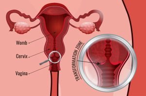
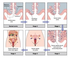
The picture shows transformation zone of the cervix. Most cervical cancers start from here.
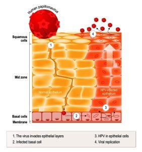
HPV virus is the most common cause of cervical cancer. The graphic shows life cycle of HPV in the human epithelium. Human papillomavirus infection which causes warts and cervical cancer (carcinoma of Cervix or malignant neoplasm) arises from the HPV infected epithelial cells.
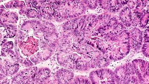
Photomicrograph of cancer cells in a biopsy from a female patient with abnormal bleeding
The image shows malignant neoplasm arising from cells in the cervix.
A biopsy is a medical test commonly performed to extract a sample of cells or tissues for examination to determine the presence or extent of the cancer. It is the only conclusive test for diagnosing and determining extent of cancer.
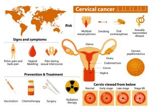
Get the facts quickly with this infographic on cervical cancer symptoms, treatment, stages, and causes (HPV or human papillomavirus).

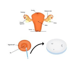
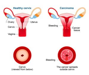
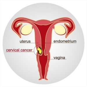
Uterine cervix is ther lowest region of the uterus. It attaches the uterus to the vagina and provides a passage between the vaginal cavity and the uterine cavity. The cervix is about 4 centimetres (1.6 inches) long, and extends about 2 centimetres into the upper vaginal cavity.
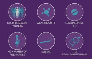
Causes and risk factors for cervical cancer include human papillomavirus (HPV) infection, having multiple sexual partners, smoking, use of birth control pills, early full-term pregnancies, chalamydia infection etc.
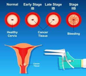
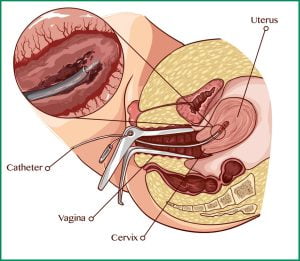
The image shows cervical cancer screening and testing by using a vaginal speculum to open the vagina. Speculum is a small tool used by the doctors dueing extracting sample or viewing the inside of the vaginal cavity.
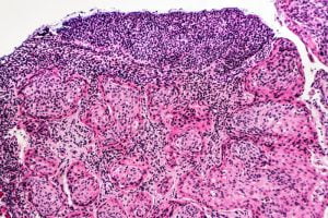
The photos shows invasive squamous carcinoma arising in association with severe squamous dysplasia, related to HPV (human papilloma virus).

Anatomically, female’s internal reproductive organs are the vagina, uterus, fallopian tubes, cervix, and ovary. The main external structures of the female reproductive system include labia majora, labia minora, clitoris, Bartholin’s glands. Cervical cancer is the cancer of cervix.
In the beginning, the cancer is diagnosed only by viewing cervical cells under a microscope. Lymph nodes are not involved at this stage. In Stage IB, the doctor can see the cancerous cells, but the cancer is found only in the cervix. In later stages, it spreads beyond the original site.
Cervical cancer is a cancer that starts from the cervix. It occurs due to the abnormal growth of cells in the cervix that have the ability to invade or spread to other parts of the body. It affects the entrance to the uterus.
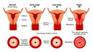
Uterine cervix is ther lowest region of the uterus. It attaches the uterus to the vagina and provides a passage between the vaginal cavity and the uterine cavity. The cervix is about 4 centimetres (1.6 inches) long, and extends about 2 centimetres into the upper vaginal cavity. In Stage IA, the cancer is diagnosed only by viewing cervical cells under a microscope. No lymph nodes are involved at this stage. In Stage IB, the doctor can see the lesion, and the cancer is found only in the cervix. In later stages, it spreads beyond the original site.
ADVERTISEMENT