


Browsing: Endometrial cancer Graphics
Comprehensive Information, Resources, and Support on Endometrial Cancer
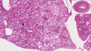
The image shows high and low power microscopic view of endometrium (uterus) in case of an endometrial cancer.

Endometrial cancer is a disease in which malignant (cancer) cells develop in the tissues of the endometrium. The endometrium is the lining of the uterus.
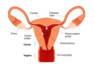
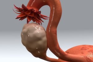
The image shows uterine polyps, which is considered as a risk factor for endometrial and uterine cancer. Your doctor will perform diagnosis to see if the polyps are cancerous.
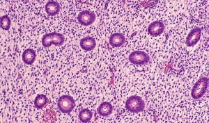
This is a photomicrograph of an endometrial biopsy in a patient with abnormal bleeding. The image shows benign proliferative endometrium, part of the normal menstrual cycle. No hyperplasia or malignancy (endometrial cancer) is seen in this case.
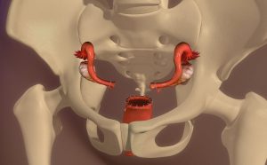
Hysterectomy is the surgical removal of uterus of women. It may also involve removal of neaby organs such as the cervix, ovaries, fallopian tubes and other surrounding structures. The image shows after hysterectomy has been performed and there is no uterus in the pelvis.
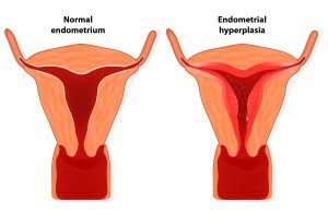
Hyperplasia is the enlargement of an organ or tissue due to an increase in the reproduction rate of its cells. This is generally considered as an initial stage in the development of endometrial cancer. If you are at high risk of cancer, your doctor will look for any signs of hyperplasia too among other signs.

Endometrial cancer signs, symptoms, and treatment
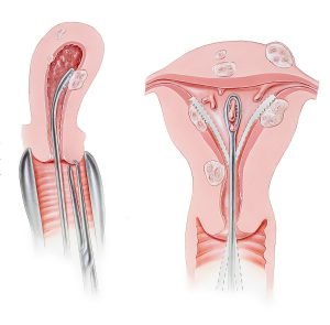
Dilation and curettage (D&C) is a brief surgical procedure to remove tissue from inside the uterus. Doctors perform this procedure to diagnose and treat certain uterine conditions such as heavy bleeding, fibroids, endometrial cancer, or to clear the uterine lining after a miscarriage or abortion.
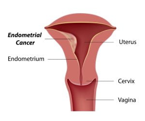
Endometrial cancer starts when cells in the inner lining of the uterus (endometrium) begin to grow out of control. It is one of the most common cancers in women in the United States.
The image shows the uterus of women with endometrial tissue as a 3D model. Endometrial cancer is the cancer of endometrial lining within the uterus.
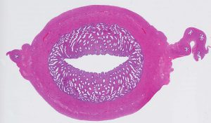
The image shows a microscopic view of the uterus with endometrium and myometrium. Endometrium is the mucous membrane that lines the inside of the uterus (womb) in women. Endometrial cancer starts from here. The myometrium is the middle layer of the uterine wall, that consists mainly of uterine smooth muscle cells.
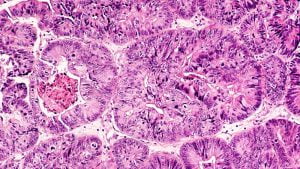
This is a photograph of adenocarcinoma of endometrium in a biopsy from a patient with abnormal bleeding. Biopsy is the ultimate test to diagnose the presence of cancer.
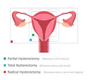
Hysterectomy is the surgical removal of uterus of women. It may also involve removal of neaby organs. The illustration shows types of hysterectomy and the portions that are rmeoved in each type of surgery.
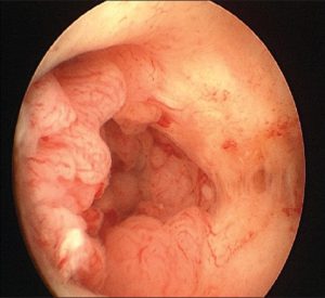
Visualization of uterus through hysterescopy shows endometrial cancer. Hysteroscopy is a procedure that allows doctors to see inside the uterus to diagnose and treat causes of abnormal bleeding. Hysteroscopy is done using a thin, lighted tube called hysteroscope that is inserted into the vagina.

Endometriosis is a risk factor for endometrial cancer. The image shows structure of the pelvic organs in a healthy woman vs in a woman with endometriosis.
ADVERTISEMENT