


Browsing: Ventricular Septal Defect Graphics
Comprehensive Information, Resources, and Support on Ventricular Septal Defect
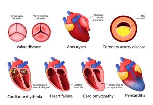
Heart disease is a term that covers a range of disorders that affect the heart. Coronary artery disease is the most common type of heart disease.
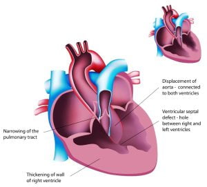
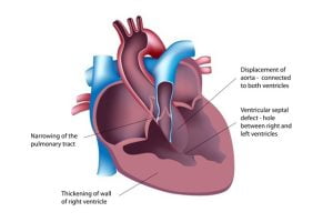
Tetralogy of Fallot (TOF) is a congenital heart defect which can cause death if left untreated. The defect occurs due to four specific reasons which are: a hole between the right and left ventricles, known as ventricular septal defect (VSD), an obstruction or narrowing in pulmonary outflow tract (connects the heart with the lungs), thickening of right ventricle, an aorta lays over the VSD and thickens. Common symptoms of TOF in children are bluish coloration of the skin caused by blood low in oxygen (cyanosis), shortness of breath and rapid breathing, fainting, heart murmur, prolonged crying in children, etc.
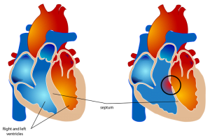
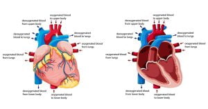
Normally, the left side of human heart only pumps blood to the body, and the right side of our heart only pumps blood to the lungs. In a person with VSD condition, there is a hole between the two ventricals. Blood can travel across the hole from the left pumping chamber (left ventricle) to the right pumping chamber (right ventricle) and out into the lung arteries. This allows oxygen-rich and oxygen-poor blood to mix together. A normal heart is shown on the left in the image and a heart with the hole is shown in the right. [Image author: Mariana Ruiz LadyofHats]
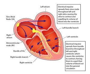
The electrical conduction system of our is responsible for sending signals generated by the sinoatrial node to cause contraction of the heart muscle. The signal generated in the sinoatrial node travels through the right atrium to the atrioventricular node, along the Bundle of His and through bundle branches to cause contraction of the heart muscle. The lower two chambers shown in the image are called left ventricle and right ventricle. They are separated by tissues. In a VSD (hole in the heart), an opening remains between the two ventricles which causes the blood to mix from both the chambers. The opening is fixed with surgery. VSD is a congenital disease that occurs in children mostly.
Our heart is a muscular organ of the size of a closed fist that functions as our body’s circulatory pump. It takes in deoxygenated blood through the veins and delivers it to the lungs for oxygenation and then pumps this oxygenated blood into the arteries. The arteries, that are part of our circulatory system, provide oxygen and nutrients to the tissues of the body by transporting the blood throughout the body. If you keep your right hand in the central part of your chest, and move it a little to the left, you can note the exact location of your your heart. The heart as four chambers.
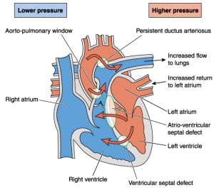
The image shows congenital heart defects resulting in abnormal blood flow through the heart and to the lungs, leading to Eisenmengers syndrome. When the heart or blood vessels near the heart do not develop normally before birth, a condition called congenital heart defect occurs. The main types of congenital heart defects are septal (atrial or ventricular) defects, cyanotic or obstructive defects. Ventricular septal defect (VSD) is a common heart defect with a hole in the heart at birth (congenital). The hole is present in the wall (ventricular septum) which separates the heart’s lower chambers as right and left ventricle. This allows the blood to pass from the left to the right side of the heart. Due to this, the oxygen-rich blood gets pumped back to the lungs instead of out to the body which makes the heart to work harder.
Low oxygen blood from the body enters the right side of the heart through two veins, the superior vena cava and the inferior vena cava. The pulmonary vein empties oxygen-rich blood from the lungs into the left atrium. Blood in the right atrium flows through right atrioventricular valve to right ventricle which results in contraction. This opens the pulmonary valve where blood flows into the pulmonary trunk. Distribution of blood by right and left pulmonary arteries to lungs unloads carbon dioxide and loads oxygen. Blood returns from the lungs through pulmonary veins to left atrium and flows through left atrioventicular valve into left ventricle. Contraction of the left ventricle opens aortic valve and blood flows into ascending aorta. Blood in aorta is distributed in the body, where it unloads oxygen and loads carbon dioxide. Blood returns to the right atrium via venae cavae. A ventricular septal defect (VSD) is a hole in septum (wall) that separates the ventricles. Due to VSD, there is an abnormal blood flow across the hole from the left ventricle to the right ventricle due to pressure difference.
ADVERTISEMENT