Dynamic Phase CT (DPCT) in Cancer Diagnosis and Prognosis
- Updated on: Sep 23, 2025
- 3 min Read
- Published on Sep 23, 2025

Understanding Dynamic Phase CT (DPCT) in Cancer Care
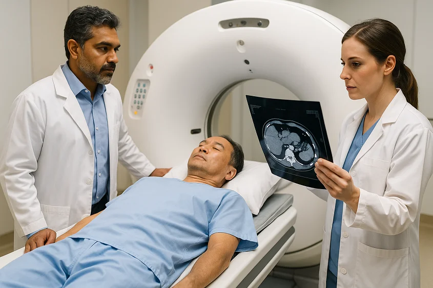
When cancer comes back after surgery or treatment, doctors often rely on advanced imaging to decide the next steps. One such imaging tool is the Dynamic Phase CT (DPCT) scan, which plays a crucial role in detecting and planning treatment for recurrent cancers. Platforms like Studycast can enhance the utility of DPCT by enabling secure sharing and collaborative interpretation of these diagnostic images. This article explains what DPCT is, why it matters, and how it affects treatment option, especially for cancers near the porta hepatis, a critical junction in the liver.
What is a DPCT Scan?
A Dynamic Phase CT scan is a special type of CT imaging done after injecting a contrast dye. Instead of taking just one set of images, the machine captures pictures in multiple phases:
– Arterial phase: right after the dye enters arteries
– Portal venous phase: when veins and the liver are filled
– Delayed phase: a few minutes later, to check how the tumor retains or releases contrast
These phases help doctors study how a tumor behaves, how close it is to blood vessels, and whether it can be surgically removed.
Why Doctors Recommend DPCT
Routine CT or PET-CT scans show if cancer has spread or how metabolically active it is. But when a tumor is very small (for example, about 1 cm) and located near vital structures like the portal vein and hepatic artery, routine scans may not provide enough clarity. That’s where DPCT helps.
|
The Porta Hepatis Challenge
The porta hepatis is the entry point into the liver where three vital structures meet:
– Portal vein
– Hepatic artery
– Bile ducts
A tumor in this area, even if just 1 cm, can cause major problems. It may block bile flow (leading to jaundice), press on blood supply, or make surgery very risky. Doctors use DPCT to check if the tumor is only touching these vessels or completely wrapping around them. This difference determines whether surgery is possible.
Surgery vs. Non-Surgery Decisions
Based on DPCT findings:
1. If the tumor is separate from vessels → surgery may be possible.
2. If the tumor is touching but not encasing → surgery with reconstruction may still be an option in specialized centers.
3. If the tumor is encasing major vessels → surgery is usually not possible.
Why Prognosis is Still Poor
Even if surgery is possible, doctors often warn patients that the overall prognosis is poor. Here’s why:
– The location limits how much tissue can be safely removed.
– Gallbladder and periampullary cancers are biologically aggressive.
– Microscopic spread may already be present even if scans look clear.
– Recurrence after surgery is common.
What Patients Can Do
Hearing ‘poor prognosis’ is never easy. But there are steps patients and families can take:
– Seek a second opinion at a hepatobiliary or GI oncology center.
– Ask about clinical trials or newer targeted therapies.
– Consider palliative surgery or stenting to relieve jaundice and improve quality of life.
– Focus on nutrition, infection prevention, and supportive care to stay strong for any treatment ahead.
When to See a Doctor?
If you have a history of gallbladder, pancreatic, or periampullary cancer and experience new symptoms like:
– Yellowing of eyes/skin (jaundice)
– Persistent abdominal pain
– Unexplained weight loss
– Itchy skin or dark urine
…do not delay. Ask your doctor about advanced imaging like DPCT to evaluate recurrence.
Final Thoughts
A Dynamic Phase CT scan is not just another test, it’s a decision-making tool. It helps doctors answer the most critical question: Is surgery possible, or do we need to focus on other treatments? While the prognosis for porta hepatis recurrence is challenging, being informed empowers patients and families to make the best choices for care.
👉 Have you or your loved one undergone a DPCT scan? Share your experience in the comments or ask your questions. We’d love to hear your story and support you.


