The field of neuroscience relies heavily on specific molecular tools to investigate the complex architecture and function of the nervous system. One such tool is the anti-rat calretinin polyclonal antibody, a reagent that plays a pivotal role in identifying and mapping neurons in the brain and spinal cord.
Calretinin, a calcium-binding protein, serves as a neurochemical marker that provides insight into neuronal subtypes and their roles in various physiological and pathological processes. This article explores the applications, mechanisms, and impact of anti-rat calretinin polyclonal antibody in neuroscience research.
Understanding Calretinin: A Brief Overview
Calretinin is a 29-kDa calcium-binding protein belonging to the EF-hand family, similar to calbindin and parvalbumin. It plays a crucial role in regulating intracellular calcium levels, which in turn influence neuronal excitability, neurotransmitter release, and synaptic plasticity. First discovered in the retina, calretinin has since been identified in various regions of the central and peripheral nervous system.
In rats, calretinin is prominently expressed in specific subsets of neurons, including interneurons in the cerebral cortex, hippocampus, cerebellum, and sensory ganglia. Because of its restricted and consistent expression patterns, calretinin is a valuable marker for delineating neuronal circuits and understanding functional neuroanatomy.
What is Anti-Rat Calretinin Polyclonal Antibody?
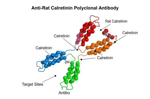
The anti-rat calretinin polyclonal antibody is generated by immunizing animals—typically rabbits or goats—with purified calretinin or its peptide sequences derived from rats. The resulting antibody pool consists of multiple immunoglobulins that recognize different epitopes on the calretinin protein, enhancing sensitivity and binding efficiency.
Polyclonal antibodies differ from monoclonal antibodies in that they target multiple sites on an antigen, making them more robust in detecting target proteins, especially under varying experimental conditions. This versatility makes the anti-rat calretinin polyclonal antibody an essential tool in both basic and applied neuroscience research.
Immunohistochemistry: Mapping the Brain
One of the most significant applications of anti-rat calretinin polyclonal antibody is in immunohistochemistry (IHC). This technique involves staining tissue sections with the antibody to visualize the distribution of calretinin-expressing neurons.
In the rat brain, calretinin-positive interneurons can be distinguished from other GABAergic interneurons by their unique morphological and electrophysiological characteristics. Using this antibody, researchers have mapped these interneurons in the hippocampus, where they play key roles in inhibitory signaling and the modulation of neural circuits.
Moreover, in the cerebral cortex, the antibody helps differentiate calretinin-positive neurons from other interneuron types like those expressing parvalbumin or somatostatin. These studies contribute to our understanding of the functional diversity among cortical interneurons, which is essential for decoding higher-order processes such as learning, memory, and cognition.
Developmental Neuroscience and Neurogenesis
Anti-rat calretinin polyclonal antibody is also instrumental in developmental neuroscience. During embryonic and postnatal development, calretinin expression serves as a marker of maturing neuronal populations. By applying this antibody to developing rat brains, researchers can trace the differentiation pathways and migration patterns of neurons.
Calretinin expression often appears early during neurogenesis and is transient in certain populations, marking critical developmental windows. Its presence is used to study the formation of sensory maps, neural circuits, and the laminar organization of the cortex.
In adult neurogenesis, particularly within the hippocampus, calretinin serves as a marker of newly generated granule cells. The antibody helps quantify neurogenic activity under different physiological conditions or in response to external stimuli such as enriched environments, stress, or pharmacological treatments.
Applications in Neurodegenerative Disease Research
Neurodegenerative diseases such as Alzheimer’s, Parkinson’s, and Huntington’s diseases are characterized by selective neuronal loss and synaptic dysfunction. Calretinin-expressing interneurons have shown varied vulnerability in different disease models, and their study offers insights into disease progression and potential therapeutic targets.
In Alzheimer’s models, for example, calretinin-positive neurons in the hippocampus and entorhinal cortex may exhibit resistance to amyloid-beta toxicity, suggesting a neuroprotective role. By using anti-rat calretinin polyclonal antibody in such studies, researchers can investigate the molecular mechanisms underlying this resistance and how calretinin might influence intracellular calcium buffering.
In Parkinson’s disease, changes in calretinin expression levels in basal ganglia structures can help decipher alterations in inhibitory circuitry. Tracking these changes through immunostaining provides valuable data for understanding disease mechanisms and assessing the impact of experimental treatments.
Studying Sensory Systems
Calretinin is prominently expressed in neurons involved in sensory processing. In the auditory system, for instance, it marks specific neuron types in the cochlear nuclei and auditory brainstem. The anti-rat calretinin polyclonal antibody is widely used to study the structural and functional organization of these sensory pathways.
In the visual system, calretinin helps identify amacrine and ganglion cell subtypes in the retina. By staining retinal sections, researchers can explore the development of visual circuits, synaptic interactions, and changes due to retinal degeneration or injury.
Similarly, in the somatosensory system, calretinin expression patterns in the dorsal root ganglia and spinal cord dorsal horn provide a window into pain processing and tactile perception mechanisms. The antibody enables detailed mapping of sensory neurons and helps identify changes due to injury or inflammation.
Calretinin as a Marker in Tumor and Lesion Studies
Though primarily used in neurobiology, anti-rat calretinin antibodies have also found relevance in neuropathology and oncology. Certain tumors, such as mesotheliomas and neuroendocrine tumors, exhibit calretinin expression. While this application is more common in human medicine, the antibody’s use in rat models aids in understanding tumor biology and testing experimental treatments in preclinical studies.
In studies involving brain lesions or trauma, researchers use the antibody to monitor the survival and reorganization of calretinin-positive interneurons. Such observations provide crucial data on neuronal plasticity and the capacity of the brain to recover or compensate following injury.
Technical Considerations and Limitations
While the anti-rat calretinin polyclonal antibody offers high sensitivity, certain technical considerations must be addressed for optimal results. Tissue fixation methods, antibody concentration, and specificity controls are critical factors that influence staining quality. Cross-reactivity with other calcium-binding proteins, although generally low, must be accounted for through proper validation.
Batch-to-batch variability in polyclonal antibodies can also affect experimental consistency. Researchers often validate new antibody lots and use internal controls to ensure reliability. Despite these limitations, the robustness of polyclonal antibodies in recognizing multiple epitopes often outweighs the challenges.
Conclusion
The anti-rat calretinin polyclonal antibody has proven to be an indispensable tool in neuroscience research. Its ability to specifically and sensitively label calretinin-expressing neurons allows researchers to explore neuronal diversity, brain development, and disease mechanisms with precision. From mapping cortical circuits and studying neurogenesis to understanding sensory processing and pathology, this antibody continues to contribute profoundly to our understanding of the nervous system.
As neuroscience advances toward more integrative and high-resolution approaches, the anti-rat calretinin polyclonal antibody will remain a foundational element in the toolbox of neurobiologists seeking to unravel the mysteries of the brain.


