


Browsing: Arrhythmia Graphics
Comprehensive Information, Resources, and Support on Arrhythmia
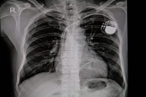
Having a pacemaker implanted is a straightforward process. It’s generally performed under local anaesthetic, and you’ll be awake during the procedure. The signal generator is usually placed under the skin near the collarbone on the left side of the chest.
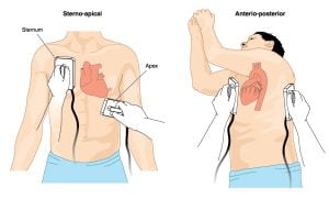
Image shows placement of defibrillator electrode paddles to perform cardioversion on a patient with cardiac arrhythmia. Cardioversion is a medical procedure that is used to restore a normal heart rhythm in people with certain types of abnormal heartbeats (called arrhythmias). Cardioversion is generally done by sending electric shocks to the heart through electrodes tied to your chest.
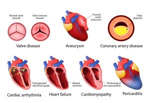
Heart disease is a term that covers a range of disorders that affect the heart. Coronary artery disease is the most common type of heart disease.
Heart arrhythmia, also known as irregular heartbeat or cardiac dysrhythmia, is a group of conditions where the heartbeat is irregular – too slow or too fast. Arrhythmias are classified into various types such as slow heartbeat (called bradycardia) and fast heartbeat (called tachycardia), and irregular heartbeat (called flutter or fibrillation).
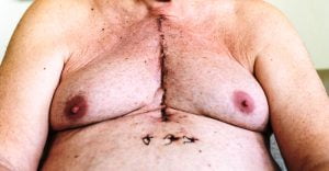
An image of a patient taken 12 days after open heart surgery. The picture shows scars where the sternum was cut in two parts, and the rib cage sprung. Just below the scar, there are holes that show locations where the drains and pacemaker cables emerged.
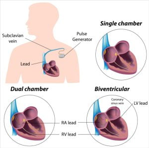
A pacemaker insertion is the implantation of a pacemaker into the chest just below the collarbone to regulate your electrical problems of the heart. It is done under the effect of a sedative and a local anesthetic at the insertion site.
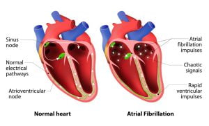
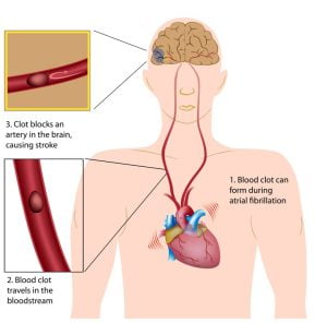
Atrial fibrillation is an irregular rapid heart rate that can increase your risk of heart-related complications. During atrial fibrillation, your heart’s two upper chambers beat out of coordination with the two lower chambers.
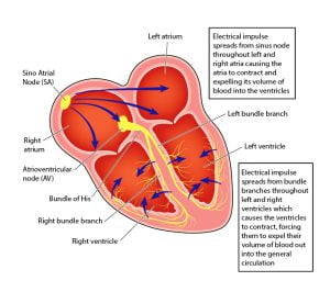
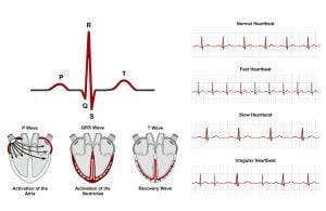
he electrical conduction system of the heart transmits signals generated by the sinoatrial node to cause contraction of the heart muscle. The signal generated in the sinoatrial node travels through the right atrium to the atrioventricular node, along the Bundle of His and through bundle branches to cause contraction of the heart muscle.
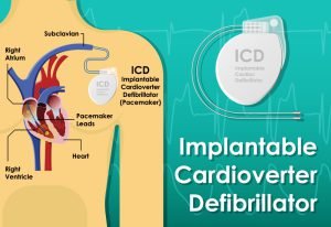
Implantable cardioverter defibrillator (ICD) consist of a pulse generator, pacing or sensing electrodes, and defibrillation coils. Its function is similar to a pacemaker. An ICD is a battery-powered device placed under the skin to monitor your heart rate. It includes wires that connect the ICD to your heart. If it detects an abnormal heart rhythm, an electric shock is sent to restore a normal heartbeat.
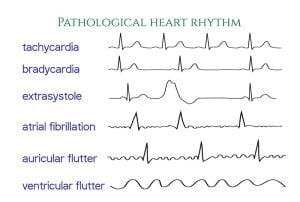
An ECG is a visual representation of the heartbeat. It can help doctors diagnose heart disorders, such as arrhythmia and other disturbances. The image shows electrocardiograms indicative of different types of irregular heart rhythms.
ADVERTISEMENT