Breast Ultrasound
- Updated on: Jul 10, 2024
- 5 min Read
- Published on Feb 18, 2019


What is a breast ultrasound?
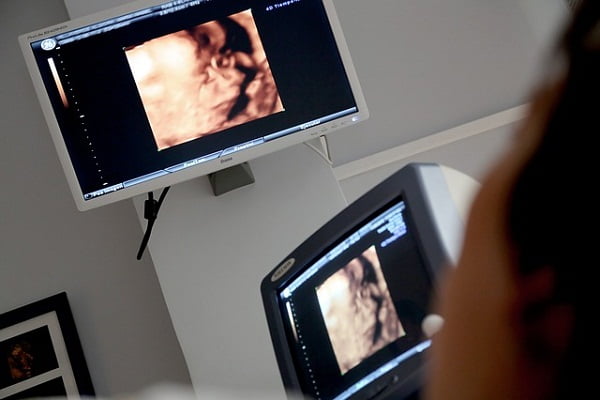
A breast ultrasound is a painless imaging technique or a diagnostic tool that uses high-frequency sound waves to produce detailed images of the inside of your breasts. During the procedure the sound waves bounce off surfaces inside your breasts and the echoes are recorded and processed into photographs or videos. It is commonly used to screen for breast abnormalities such as undefined breast mass and tumors. Ultrasounds do not use radiations and hence is considered safe for pregnant woman and breast feeding mothers as compared to an X-ray and an MRI scan. It is also used to see the blood flow in different areas of your breasts.
Breast ultrasound is performed very commonly after a mammogram to tell whether a lump in a breast is a cyst (a fluid-filled sac) or a solid mass, which might be breast cancer. It is also helpful revealing the location of a tumor in your breast and this can help the doctors to decide the exact place to insert a needle during a biopsy. Sometimes breast ultrasound can also find out if a nodule recorded on a mammogram is either a solid nodule or a cystic lesion.
Purpose of a breast ultrasound: Why do you need a breast ultrasound?
There are many reasons and purposes of why a breast ultrasound may be recommended. Your doctor may perform a breast ultrasound as an initial or early diagnostic tool to evaluate breast lumps. Since it doesn’t use any ionizing radiation, it is often recommended for individuals who are not an ideal candidate for radiation bases imaging techniques. A breast ultrasound also allows your doctor to determine the location and size of the lump.
You may have to go through this procedure while your doctor is performing biopsy of your breast lump. Here, an ultrasound is used as guide to reach the lump and collect the tissues from it. This procedure is known as ultrasound guided biopsy.
Ultrasound is useful for looking at breast lumps especially those that can be felt but not seen on a mammogram. Mammogram is not able to capture the lumps in women with dense breast tissue. It can also be used to aid in visualisation of changes seen on a mammogram.
Apart from being used to determine the nature and extent of a breast abnormality, a breast ultrasound may also be recommended for women who should avoid radiation, such as women under age 25, pregnant women, breast feeding women and women with silicone breast implants.
Other various reasons for which a breast ultrasound may be performed include:
- in case of dense breast tissue when a mammogram may not be able to see through the tissue
- if you are having unusual nipple discharge, which may be a sign of breast cancer or other breast problem
- if you are feeling mastitis or inflammation of the mammary tissues
- for breast implants evaluation
- to assess symptoms, such as breast pain, redness, and swelling
- to assess discoloration or changes in your breast skin
- monitoring of existing benign breast lumps
Can ultrasound detect cancer?
Ultrasound can’t detect breast cancer on its own neither it is used as screening test for breast cancer. It is rather used to complement and aid other screening tests. If your doctor felt any abnormality during a physical exam or if any deviations are seen on a mammograph, ultrasound is considered the best way to find out if the abnormality is solid as in case of a benign fibroadenoma or cancer or fluid-filled such as a benign cyst. It cannot confirm if the solid lump is cancerous or if any calcification is present.
If you are aged less than 30 years, your doctor may recommend ultrasound before mammography to evaluate a breast lump that can be felt through the skin also known as palpable lump. Usually women at younger age tend to have dense breast tissue and full of milk glands, mammograms can be difficult to interpret for cancer in these women as these glandular tissues look dense and white very much like a cancerous tumor.
Breast ultrasound procedure: How should you prepare for breast ultrasound?
You may have your breast ultrasound as an outpatient basis or as a part of your stay in the hospital. The way this breast ultrasound procedure is performed may vary depending on your healthcare provider’s practices and your condition.
Generally, breast ultrasound procedure consists of the following consecutive steps:
- First you will be asked to remove clothing and any jewellery from the waist up and will be provided a gown to wear.
- You will be asked to lie on your back on an exam table. The technologist will instruct you to raise your arm above your head on the side of the breast to be examined. It might be possible that you will be asked to lie down on your side.
- A clear and warm gel will be applied on the skin over the breast area to be looked at by the technologist.
- Next the technologist will press the transducer against the skin and move it over the area being studied.
- Once the test procedure is finished, the technologist will wipe off the gel.
How to read a breast ultrasound?
Usually breast ultrasound images are analyzed by a radiologist. Radiologist is a physician specifically trained to interpret and supervise radiology examinations. He will then prepare a signed report based on his analysis of ultrasound images and will send to your primary care physician, or to the physician or other healthcare provider who requested the exam. Your referring physician will share the results with you in simple terms which you can understand. Sometime the radiologist may also discuss the results with you after your examination has been concluded.
Breast ultrasound results: What does a normal breast ultrasound mean?
The images received after breast ultrasounds are in black and white. Abnormal growths, cysts, and tumors will appear as dark areas on the scan. However you should not interpret a dark spot on your ultrasound as a breast cancer. A confirmed diagnosis can be made by your healthcare provider after examining and diagnosing different virtues of an abnormality seen on the scan. Read about breast cancer diagnosis.
As a fact, majority of breast lumps are benign. The cause of these benign lumps in the breast can vary which may include:
- An adenofibroma – benign tumor of the breast tissue
- Fibrocystic breasts – breasts that are lumpy and painful because of hormonal changes
- An intraductal papilloma – small, benign tumor of the milk duct
- Mammary fat necrosis – bruised, dead, or injured fat tissue
If your doctor finds any lumps on the ultrasound scan, he/she will first perform a breast MRI and then will go for breast biopsy of the lump to confirm whether the lump is cancerous or benign. If nothing abnormal is seen on the ultrasound image screen, it means your breast ultrasound is normal.


