



How Ultrasound Technology Has Transformed Modern Healthcare
- Updated on: Aug 12, 2026
- 4 min Read

Dr. Rida Maryam is a dedicated medical practitioner with a focus on the management of chronic diseases. Her areas of interests include diabetes and women health. Dr. Maryam’s commitment to her specialty is rooted in a strong academic background and a deep passion for improving delivering quality and passionate care to her patients.
- Published on Aug 12, 2026
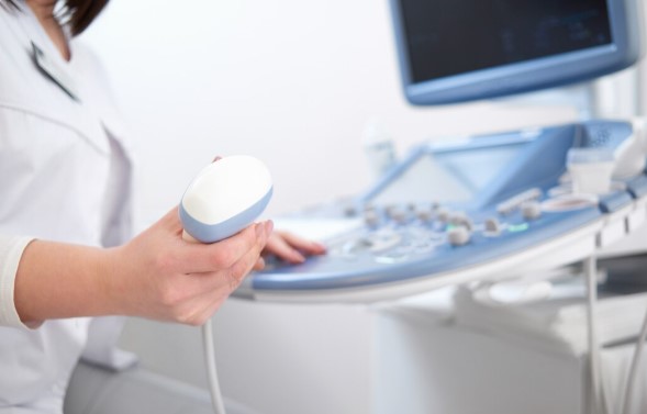
Ask most people what ultrasound is for, and they’ll say pregnancy. That answer was reasonable forty years ago. Today it describes maybe a fraction of how the technology actually gets used, and the gap between public perception and clinical reality is wider here than with almost any other imaging tool in medicine.
One characteristic explains much of that expansion. As the FDA describes it, sonography images are captured in real time and can show movement of internal organs as well as blood flowing through vessels, and unlike X-ray imaging, there is no ionizing radiation exposure involved. Patients across Austin encounter this technology far more often than they realize.
ADVERTISEMENT
ADVERTISEMENT
Here’s exactly how it reshaped diagnosis: the specific ways ultrasound technology changed
No Radiation Makes It Uniquely Repeatable
Every imaging decision involving CT or X-ray weighs diagnostic benefit against radiation exposure, and that calculation limits how often those studies can reasonably be repeated. Procedures like sonography sidestep the question entirely because they use sound waves rather than ionizing radiation. This distinction alone opens up monitoring approaches that simply aren’t practical with radiation-based imaging, regardless of how valuable repeated scanning might otherwise be.
This matters most in situations requiring repeated monitoring, tracking a known finding over months, or following a condition through treatment. It also makes this particular technique the default for imaging in pregnancy and in children, where cumulative radiation exposure is a genuine concern. That said, the FDA notes ultrasound energy can produce biological effects, including slight tissue heating, which is why prudent use by trained operators still matters.
Real-Time Imaging Guides Procedures as They Happen
Static images tell you what something looks like. Real-time imaging tells you what it’s doing and lets a clinician watch their own instrument move through tissue as it happens. That capability changed procedural medicine substantially.
- Biopsies: needle placement guided directly by live imaging rather than anatomical estimation alone
- Vascular access: locating and accessing veins or arteries with visual confirmation in real time
- Joint injections: placing medication precisely within a joint space under direct visualization
- Drainage procedures: guiding catheter or needle placement for fluid removal with continuous imaging feedback
The accuracy improvement is meaningful, and the technique has become standard practice across specialties that previously relied on feel and estimation. Patients scheduled for an ultrasound in Austin may be having it for diagnostic imaging or as guidance for a separate procedure, and the two involve different preparation. Longhorn Imaging includes ultrasound among the modalities it offers, and confirming which type of study has been ordered is worth doing when scheduling.
Portability Brought Imaging to the Bedside
This technique is the only major imaging modality that can travel to the patient rather than requiring the patient to travel to it. Handheld and cart-based units now appear in emergency departments, intensive care units, ambulances, and rural clinics. This mobility fundamentally changes the logistics of care, since a critically ill patient no longer has to be stable enough to be moved before imaging can even begin.
This shifted where certain decisions get made. A clinician can assess for internal bleeding, cardiac function, or fluid in the lungs within minutes of a patient arriving, without transport to a radiology suite. In time-critical situations that difference genuinely affects outcomes, and in resource-limited settings it may represent the only imaging available at all
Applications Now Reach Far Beyond Pregnancy
Obstetric imaging remains a major use, but ultrasound now covers a considerably broader range of clinical applications.
- Cardiac echocardiography: assessing heart structure, valve function, and pumping efficiency in real time, without exposing the patient to radiation during repeated monitoring
- Vascular studies: evaluating blood flow and identifying clots within veins and arteries, often used to detect conditions like deep vein thrombosis before they become more serious
- Abdominal imaging: examining the liver, gallbladder, kidneys, and pancreas for structural abnormalities, cysts, or signs of disease that wouldn’t be visible through physical examination alone
- Thyroid and breast evaluation: assessing structural changes in soft tissue with high resolution, frequently used to characterize nodules or masses found during a physical exam
- Musculoskeletal assessment: evaluating tendons and joints for injury, inflammation, or degeneration, often guiding decisions about physical therapy versus further intervention
Each of these represents a question this technique answers well enough to be a first-line choice rather than a fallback. The breadth is what makes the pregnancy association so outdated, though it persists because that’s the context in which most people personally encounter it.
Accessibility and Cost Compared to Other Modalities
Ultrasound equipment costs substantially less than CT or MRI systems, requires less specialized facility infrastructure, and generates lower per-study costs. Those economics affect access directly, particularly outside major medical centers.
The trade-off is real, though. It is operator-dependent in a way CT and MRI aren’t, meaning image quality and diagnostic accuracy vary with the skill of the person performing the scan. It also has genuine limitations, including poor penetration through bone and air, which is why it complements rather than replaces other imaging.
What to Expect During an Ultrasound Exam
The exam itself is straightforward, though preparation varies by study type:
- Preparation instructions: some abdominal studies require fasting, while pelvic studies may require a full bladder, so confirm when scheduling
- The gel and transducer: a thin layer of gel is applied so sound waves transmit properly, and the probe is moved across the skin
- Duration and comfort: most studies take between fifteen and forty-five minutes, and the exam is generally painless
- Results timing: images are typically reviewed by a radiologist afterward rather than interpreted during the scan
Asking about preparation when booking prevents the common frustration of arriving for a study that has to be rescheduled.
Conclusion
Ultrasound rarely gets described as revolutionary, which is odd given how thoroughly it changed clinical practice. It made imaging repeatable without accumulating risk and cheap enough to be genuinely accessible. It turned certain procedures from landmark-guided estimation into directly visualized precision. None of that came from a single breakthrough moment, which is probably why it gets less attention than technologies with more dramatic origin stories. What it does have is a safety profile that permits use in situations where other imaging simply can’t be justified, and that alone accounts for a great deal of modern diagnostic medicine.
Share this article



Latest Posts





ADVERTISEMENT